
Archivo:HIV-budding-Color.jpg

Tamaño de esta previsualización: 800 × 531 píxeles. Otras resoluciones: 320 × 213 píxeles | 640 × 425 píxeles | 1024 × 680 píxeles | 1280 × 850 píxeles | 2967 × 1971 píxeles.
Archivo original (2967 × 1971 píxeles; tamaño de archivo: 3,92 MB; tipo MIME: image/jpeg)
| Éste es un fichero de Wikimedia Commons, un depósito de contenido libre hospedado por la Fundación Wikimedia. Más abajo se reproduce su página de descripción con la información sobre su origen y licencia. |
Resumen
| Descripción |
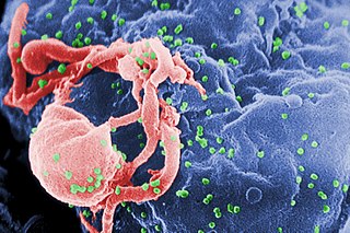
English: Scanning electron micrograph of HIV-1 budding (in green) from cultured lymphocyte. This image has been colored to highlight important features; see PHIL 1197 for original black and white view of this image.
Multiple round bumps on cell surface represent sites of assembly and budding of virions.
Español: Microfotografía con MEB de VIH-1 en liberación (en verde) en un cultivo de linfocitos. Esta imagen ha sido coloreada para resaltar las características importantes; para la imagen original en blanco y negro véase PHIL 1197. Las múltiples protuberancias redondeadas sobre la superficie celular representa los sitios de ensamblado y gemación de viriones.
Français : Virus HIV fixé sur un lymphocyte vu en microscopie électronique (fausses couleurs, le VIH est en vert).
Bahasa Indonesia: HIV yang baru memperbanyak diri tampak bermunculan sebagai bulatan-bulatan kecil (diwarnai hijau) pada permukaan limfosit setelah menyerang sel tersebut; dilihat dengan mikroskop elektron.
Русский: Фотография, полученная с помощью сканирующего электронного микроскопа. Вирусы ВИЧ (зелёные) отпочковываются от заражённого лимфоцита. Фотография была раскрашена с целью подчеркнуть важные детали; см. исходную чёрно-белую версию ниже.
Многочисленные круглые выпуклости на поверхности клетки являются местами сборки и отпочковывания вирионов.
Български: Вирусът ХИВ (в зелено) разспространяващ се от вече заразен лимфоцит.
Polski: Fotografia wykonana skaningowym mikroskopem elektronowym - przedstawia wirusy (kolor zielony) wydostających się z limfocytu. |
||
| Fecha | |||
| Fuente |
|
||
| Autor |
|
||
| Permiso (Reutilización de este archivo) |
PD-USGov-HHS-CDC English: None - This image is in the public domain and thus free of any copyright restrictions. As a matter of courtesy we request that the content provider be credited and notified in any public or private usage of this image. |
||
| Otras versiones |
|


fuk12
Licencia
Esta imagen es una obra de los Centros para el Control y la Prevención de Enfermedades, parte de los Departamento de Salud y Servicios Humanos de los Estados Unidos, adoptadas o realizados durante el desempeño de funciones oficiales de un empleado. Como una obra de los Estados Unidos del gobierno federal, la imagen es de dominio público.
|
Historial del archivo
Haz clic sobre una fecha y hora para ver el archivo tal como apareció en ese momento.
| Fecha y hora | Miniatura | Dimensiones | Usuario | Comentario | |
|---|---|---|---|---|---|
| actual | 00:16 20 abr 2008 | | 2967 × 1971 (3,92 MB) | Optigan13 | {{Information |Description={{en|Scanning electron micrograph of HIV-1 budding from cultured lymphocyte. See PHIL 1197 for a black and white view of this image. Multiple round bumps on cell surface represent sites of assembly and budding of virions.}} |Sou |
Usos del archivo
Las siguientes páginas usan este archivo:
Uso global del archivo
Las wikis siguientes utilizan este archivo:
- Uso en ar.wikipedia.org
- Uso en arz.wikipedia.org
- Uso en ast.wikipedia.org
- Uso en as.wikipedia.org
- Uso en azb.wikipedia.org
- Uso en az.wikipedia.org
- Uso en be-tarask.wikipedia.org
- Uso en bg.wikipedia.org
- Uso en bn.wikipedia.org
- Uso en ca.wikipedia.org
- Uso en ca.wikinews.org
- Uso en ckb.wikipedia.org
- Uso en cs.wikipedia.org
- Wikipedie:Studenti píší Wikipedii/Pokroky v imunologii I (2013/2014)
- Wikipedie:Studenti píší Wikipedii/Pokroky v imunologii I (2014/2015)
- Wikipedie:Nástěnka/Univerzita Karlova/Pokroky v imunologii (2013-2014)
- Wikipedie:Nástěnka/Univerzita Karlova/Molekulární imunologie (2014-2015)
- Wikipedie:Nástěnka/Univerzita Karlova/Pokroky v imunologii (2014-2015)
- Uso en cy.wikipedia.org
- Uso en de.wikipedia.org
- Uso en diq.wikipedia.org
- Uso en en.wikipedia.org
- Uso en en.wikibooks.org
Ver más uso global de este archivo.